Get Easy Health Digest™ in your inbox and don’t miss a thing when you subscribe today. Plus, get the free bonus report, Mother Nature’s Tips, Tricks and Remedies for Cholesterol, Blood Pressure & Blood Sugar as my way of saying welcome to the community!
That snap, crackle and pop in your knee may start with your thyroid
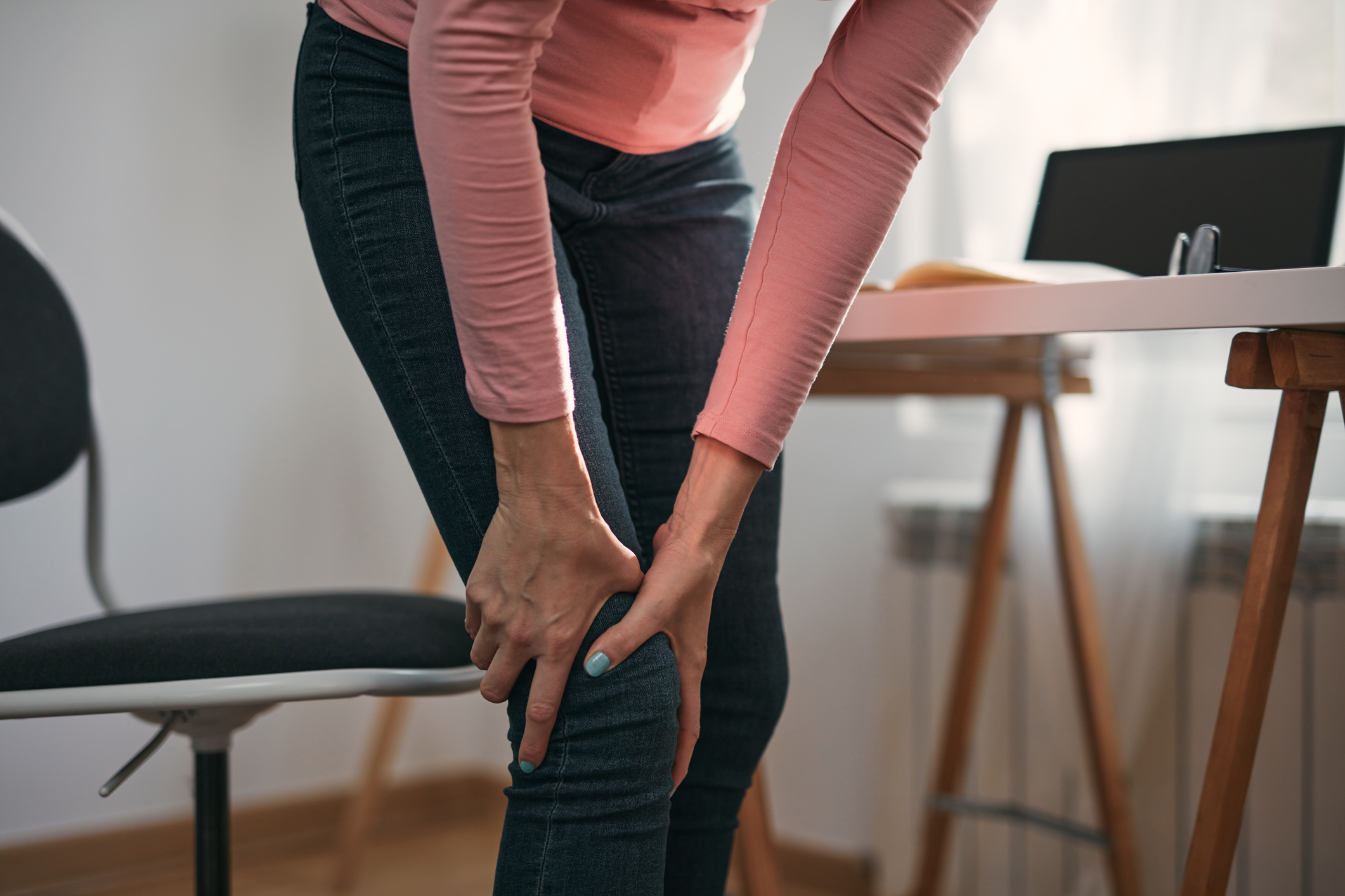
The other day I was getting up from a squat and I noticed a sort of crackling sound coming from my right knee.
It didn’t hurt, so I really didn’t think much of it. It’s a condition known as crepitus, and it usually just means there are air bubbles popping in the joint.
When air is the cause, crepitus is harmless. But I’m tempted to get my knee checked out anyway because I’ve discovered there are other causes of crepitus that aren’t as benign…
Calcium crystals can damage cartilage
Knee osteoarthritis (OA), the most common form of arthritis, affects 34 million people in the U.S., and there are no available treatments to prevent its progression. Its symptoms include pain, inflammation, swelling, instability and weakness in the joint — and a crackling sound that’s been compared to Rice Krispies.
There’s another type of arthritis that causes symptoms similar to knee OA — calcium pyrophosphate deposition (CPPD). Also known as “pseudogout,” CPPD involves the formation of calcium pyrophosphate (CPP) crystals in the blood that then settle in joint cartilage. These CPP crystal deposits trigger an inflammatory attack in the joint, causing pain, stiffness, swelling and (you guessed it) a crunching or crackling sound.
Calcium crystals can also be found in the joints of people with knee OA. Until recently, they were believed to be harmless and potentially something that happens with old age.
But U.S. researchers using computed tomography (CT) found that calcium crystal deposits in the knee can contribute to the worsening of joint damage. The researchers are the first to use computerized X-ray imaging, which are more sensitive to detecting calcium crystals than regular X-rays.
The study evaluated participants from the Multicenter Osteoarthritis (MOST) study for intra-articular mineralization (IAM) based on its location within the knee. They then examined the effect on cartilage via MRI over two years. The average age of the participants was 60.
With CT, the researchers were able to detect a higher amount of deposits than previously found by plain X-rays. Study results showed an increased risk of cartilage damage on follow-up, including in knees without any damage to begin with, supporting the theory that calcium crystal deposition in the joint was the cause.
“The cartilage damage is most likely to occur in the same locations where the crystals are deposited, suggesting a localized effect,” says corresponding author Dr. Tuhina Neogi, a professor at Boston University Chobanian & Avedisian School of Medicine.
“We have also shown that these crystals can contribute to knee pain in another recently published paper,” Neogi says. “Taken together, these findings highlight the important role of calcium crystals in structural damage and symptoms in knee osteoarthritis.”
Neogi adds that with the identification of this link, researchers can focus on identifying ways to prevent these crystal deposits from occurring with the hope of relieving pain and limiting progression of joint damage in OA.
What causes calcium crystal deposits?
While it’s not definitively known why CPP crystals form, there are theories. It’s believed excess iron or calcium, low magnesium, and an overactive or severely underactive thyroid gland may be contributing factors. A healthy functioning thyroid is important for mineral metabolism, especially bone tissue mineral density.
So if you’re looking to lessen the risk of these painful conditions, it’s a good idea to make sure your thyroid is functioning properly, your magnesium levels are optimal, and you’re not getting too much iron or calcium. You also want to check your levels of vitamins D and K2 (part of an emerging group of vitamins that fight a common contributor of unhealthy aging), both of which make sure calcium is being directed to the bones, where it’s most beneficial.
There are nutrients that support good thyroid function: iodine, copper, selenium and zinc. Iodine is particularly important, as the thyroid needs iodine to produce thyroid hormone. Some good sources of iodine include organic yogurt, cranberries, iodized salt, navy beans and sea vegetables like kelp and wakame.
Be aware that it gets more difficult for your body to absorb iodine as you get older, so you may need an iodine supplement to ensure you’re maintaining healthy levels of the nutrient. Also, you can increase thyroid hormone efficiency by combining iodine with the amino acid L-Tyrosine.
Editor’s note: Have you heard of EDTA chelation therapy? It was developed originally to remove lead and other contaminants, including heavy metals, from the body. Its uses now run the gamut from varicose veins to circulation. Click here to discover Chelation: Natural Miracle for Protecting Your Heart and Enhancing Your Health!
Sources:
Calcium crystal deposits in the knee found to contribute to joint damage — Medical Xpress
Intra-Articular Mineralization on Computerized Tomography of the Knee and Risk of Cartilage Damage: The Multicenter Osteoarthritis Study — Arthritis & Rheumatology
Calcium Pyrophosphate Deposition (CPPD) — American College of Rheumatology
Calcium Pyrophosphate Deposition — Arthritis Foundation
Knee, shoulder & elbow cracking or popping (crepitus) — Aurora Health Care
Common Knee Osteoarthritis Symptoms — American Knee Pain Centers
Snap, Crackle & Pop: Why Do My Knees Make Noises—And Should I See A Doctor? — Henry Ford Health


